Moyae's New Digital Tool Is Transforming Retina Documentation


If you're a retina specialist, you know the drill. You're in the exam room, fundus camera images pulled up, and you reach for that white piece of paper with the two circles representing the retina. You sketch the macula, note the hemorrhages, and scribble a few notes in the margin. Later, someone (maybe you) manually transcribes those markings into your EMR, cross-references them with the appropriate ICD-10 codes, and hopes nothing got lost in translation.
It's 2026. We can do better.
## The Hidden Costs of Paper Retina Drawings
What seems like a simple workflow creates multiple friction points that add up across dozens of patients daily:
- **Documentation delays**: Paper drawings sit on desks until someone has time to scan or manually enter them into patient records
- **Billing reconciliation gaps**: Translating a hand-drawn dot on a retina into the correct ICD-10 code (H35.32 vs. H35.33?) introduces human error and claim denials
- **Historical blind spots**: Flipping between scanned PDFs of past drawings to track disease progression is slow and imprecise
- **Legibility disasters**: We've all seen it—coffee stains, smudged ink, or that one doctor's handwriting that even pharmacists can't decode
Most EMRs treat retina drawings as an afterthought: a fillable form field at best, an upload button for scanned images at worst.
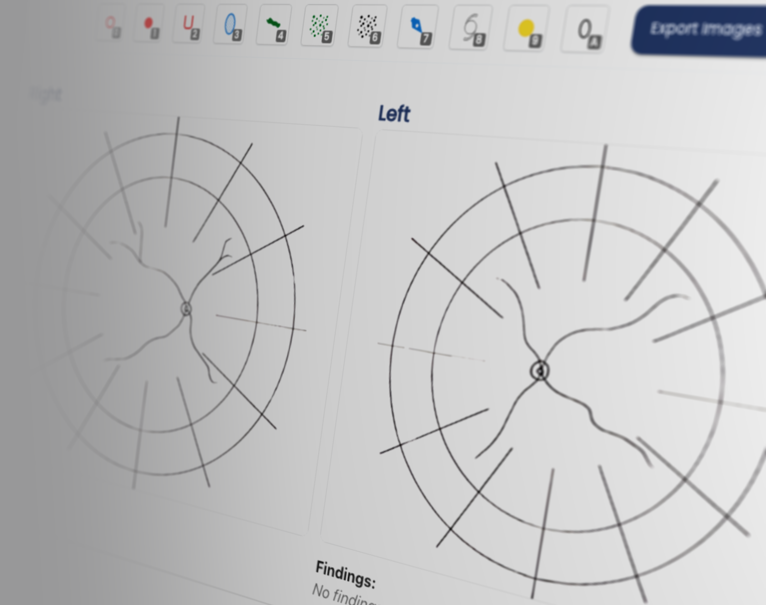
## Introducing Moyae's Retina Drawing Component
We built our new Retina Drawing tool because retina specialists told us they needed something that actually understood their workflow. Not a generic diagram tool. Not a digital canvas that happens to have an eye template. A purpose-built solution designed from day one wit
h practicing retina specialists.
### 1. Draw Once, Use Everywhere
Create detailed retina diagrams directly within the patient encounter. Mark drusen, edema, and more. We've included every annotation tool retina specialists actually use. These drawings automatically embed into:
- The encounter summary for your quick review
- Patient records for longitudinal tracking
No scanning. No uploading. No duplicate data entry.
### 2. Billing Intelligence Built-In
Each marker you place isn't just a visual annotation, it's a structured data point. When you mark a region as "macular edema," the system automatically suggests an eye-specific ICD10 code.
✅ **Automated ICD-10 Matching** – Every annotation maps to the appropriate billing code
✅ **Audit Trail Transparency** – See exactly which markings generated which codes
✅ **Reduced Claim Denials** – Structured data means cleaner submissions from the start
### 3. Designed by the Doctors Who Use It
We didn't lock engineers in a room and ask them to build a drawing tool. We watched retina specialists work. We learned how they hold their pens, when they zoom in on the macula, which findings they always circle versus shade.
The result? A tool that feels like an extension of your clinical thought process:
- **Contextual brushes** – Different tools for cotton wool spots vs. microaneurysms
- **Anatomical guides** – Smart overlays for vascular arcades, optic nerve boundaries, and macular subfields
- **One-click templates** – Pre-populate drawings for common presentations (wet AMD, PDR, BRVO)
## From Isolated Sketch to Integrated Intelligence
What makes this more than just a digital notepad is how it connects to the broader Moyae ecosystem. Your retina drawing becomes a FHIR object immediately, making it:
- **Searchable** – Find all patients with "temporal hemorrhage" across your entire practice
- **Trendable** – Visualize lesion changes over time with automated progression analysis
- **Interoperable** – Share structured drawings with referring physicians, not illegible scans
- **AI-Ready** – Future enhancements will flag concerning changes between visits automatically
## The Future of Retina Documentation
Retina specialists have one of the most visually-demanding specialties in medicine. You interpret subtle changes in OCT scans, fluorescein angiograms, and fundus photographs every day. Your documentation tools should match that precision.
With Moyae's Retina Drawing component, we're not just replacing paper, we're reimagining what's possible when documentation works for you instead of against you:
📊 **Faster encounters** – No more pausing to scan or transcribe
💰 **Cleaner billing** – Automated coding reduces administrative rework
👁️ **Better care** – Instant visual recall of previous visits improves treatment decisions
📈 **Richer data** – Structured drawings enable population health insights and research
**Ready to see your retina documentation transform?** Schedule a short demo and we'll show you how practices are already saving time per retina encounter.
📅 [Book a Demo](https://www.moyae.com)
---
*P.S. – This release is just the beginning. We're already working on OCT overlay integration and AI-powered progression detection. Stay tuned.*


.png)
.png)